|
|
Aortic Stenosis
General Considerations
- Most often as result of degeneration of bicuspid aortic valve
- Less commonly rheumatic heart disease or secondary to degeneration
of a tricuspid aortic valve in person > 65
Location
- Supravalvular
- Uncommon
- Associated with William’s Syndrome
- Hypercalcemia
- Elfin facies
- Pulmonary stenoses
- Hypoplasia of aorta
- Stenoses in
- Renal, celiac, superior mesenteric arteries
- Valvular
- Most common
- Either congenital (from a bicuspid aortic valve) or acquired
- Bicuspid aortic valve is the most common congenital cardiac anomaly
- Subvalvular
- Associated with
- Hypoplastic left heart syndrome
- Idiopathic Hypertrophic Subaortic Stenosis
- Hypertrophic cardiomyopathy
- Subaortic fibrous membrane
Types
- Congenital aortic stenosis (more common)
- Most frequent congenital heart disease associated with
intra-uterine growth retardation (IUGR)
- Subvalvular (30%)
- Valvular (70%)
- Degeneration of bicuspid valve
- Supravalvular
- Acquired aortic stenosis
- Rheumatic valvulitis
- Almost invariably associated with mitral valve disease
- Fibrocalcific senile aortic stenosis
Clinical Findings
- Asymptomatic for many years
- Classical triad
- Angina
- Syncope
- Shortness of breath (heart failure)
- Systolic ejection murmur
- Carotid pulsus parvus et tardus
- Diminished aortic component of 2nd heart sound
- Sudden death in severe stenosis after exercise
- Diminished flow in coronary arteries causes ventricular dysrhythmias
and fibrillation
- Decompensation leads to left ventricular dilatation and pulmonary
venous congestion
Imaging Findings
- In older children or young adults
- Prominent ascending aorta
- Poststenotic dilatation of ascending aorta
- Left ventricular heart configuration
- Normal-sized or enlarged left ventricle
- Concentric hypertrophy of left ventricle produces a relatively small
left ventricular chamber with thick walls
- Heart size is frequently normal
- In adults >30 years
- Prominent ascending aorta
- Poststenotic dilatation of ascending aorta
- Calcification of aortic valve (best seen on RAO)
- In females, usually indicates hemodynamically significant aortic stenosis
- Calcification of the valve usually indicates a gradient across
valve of > 50mm Hg
- Calcification begins in bicuspid and rheumatic valve in 4th decade
but not until > 65 in tricuspid
- DDx
- Calcification of aortic annulus in elderly
- Calcified coronary artery ostium (thickened cusp echoes only in diastole)
- Normal to enlarged left ventricle
Echocardiographic findings
- Thickened and calcified aortic valve with multiple dense cusp echoes
throughout cardiac cycle
- Right > non-coronary > left coronary cusp
- Decreased separation of leaflets in systole with reduced opening orifice
- (13-14 mm = mild AS; 8-12 mm = moderate AS; <8 mm = severe AS)
- ± Doming in systole
- Dilated aortic root
- Increased thickness of LV wall (= concentric LV hypertrophy)
- Hyperdynamic contraction of LV (in compensated state)
- Decreased mitral EF slope (reduced LV compliance)
- LA enlargement
- Increased aortic valve gradient (Doppler)
- Decreased aortic valve area (unreliable)
Angiographic findings
- Simultaneous LV and aortic pressures recordings yield valve gradients from left heart
catheterization
- Angiographic technique uses standard RAO left ventriculogram and an aortogram
using a 40° LAO projection
- A non-calcified, bicuspid valve reveals thickening and doming of the valve leaflets
in systole
- A jet of non-opacified blood is visible through stenotic valve
- Congenitally bicuspid valves still usually have three aortic sinuses with one large
non-coronary sinus equal in size to the other two
- Calcification begins in the bicuspid and rheumatic valve in the 4th decade
but not until >65 in tricuspid
- In rheumatic disease, the aortic valve commissures usually fuse whereas they
do not in the degenerated tricuspid valve
Differentiating Causes of Aortic Stenosis
| Etiology/Findings |
Calcification |
Other clues |
| Congenital Bicuspid Valve |
30’s |
Jet effect on aortogram |
| Degeneration of Tricuspid Valve |
> 65 |
Coronary artery ca++
Commissures don’t fuse |
| Rheumatic dz in Tricuspid Valve |
30’s here; teens in 3rd
world countries |
MS or MR almost always present;
commissures fuse |
Valve areas
Normal |
Mild |
Severe |
Critical |
2.6-3.5cm2 |
1.3-1.7 |
1.0 |
0.5 |


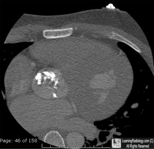
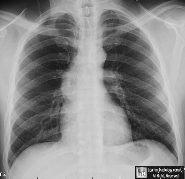
Aortic Stenosis. Top: Axial CT scan through heart demonstrates a heavily
calcified aortic valve (white arrow).
Bottom: Frontal chest radiograph in
another patient with aortic stenosis shows a dilated
ascending
aorta (white arrow) that abnormally projects farther
to the right than the right heart border.
This is caused by
post-stenotic dilatation of the aorta.
For these same photos without the arrows, click here and here
For more information, click on the link if you see this icon 
|
|
|
